Early pregnancy detection is one of the highest-return practices in cattle production. Identifying an open cow at 28 days post-breeding instead of 45 days — a difference of just 17 days — can save 3to5 per day in feed and maintenance costs for that animal while shortening the calving interval and getting her rebred faster. Over a 100-cow herd, those 17 days add up quickly. A portable veterinary ultrasound scanner makes this early detection possible, reliably and non-invasively, right in the barn or on pasture.
But how does ultrasound technology actually work for livestock? What makes a portable unit different from a cart-based clinic system? And at what point after breeding can you realistically confirm pregnancy in cattle, sheep, and other species? This guide explains the technology, the research behind early pregnancy diagnosis, and what to look for in a field-ready scanner.
What Is a Portable Veterinary Ultrasound Scanner
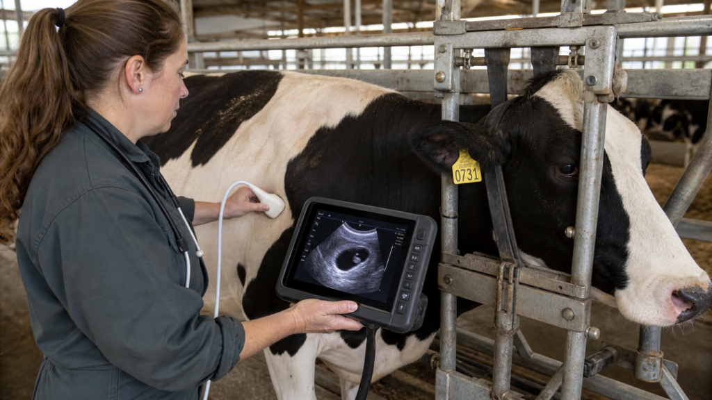
A portable veterinary ultrasound scanner is a handheld diagnostic device that uses high-frequency sound waves to create real-time images of internal reproductive organs. Unlike cart-based ultrasound machines that stay in a clinic, a portable unit weighs under one kilogram and operates on battery power for a full day of field work — moving through pens, pastures, and handling chutes without cords or carts.
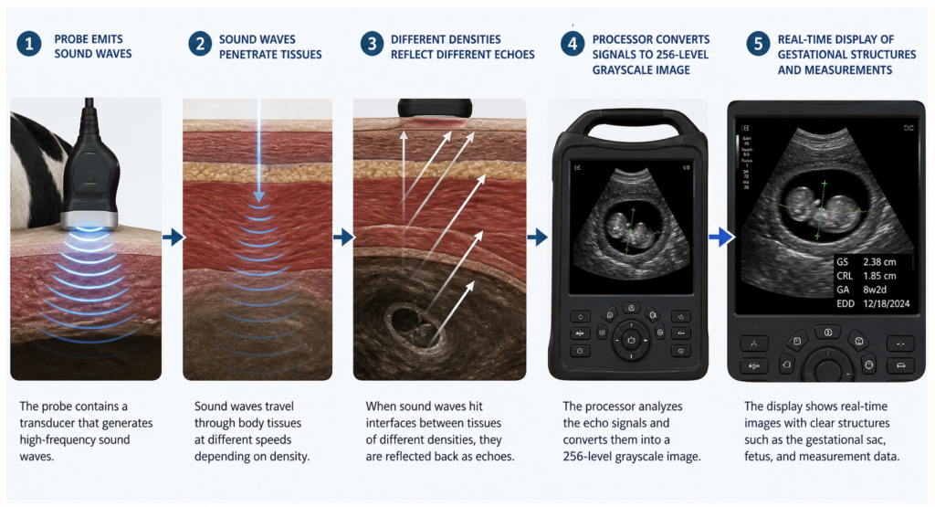
The scanner works on the same physical principle as all medical ultrasound: a probe emits high-frequency sound waves (typically 3.5 MHz for cattle) that penetrate soft tissue. When these waves encounter boundaries between different tissue types — the wall of a uterus, fluid inside a gestational sac, a fetal skeleton — some of the energy reflects back as an echo. The probe receives these echoes, and the scanner’s processor converts them into a 256-grayscale image displayed in real time on the screen.
Scientific literature confirms that ultrasound is widely used for pregnancy diagnosis in livestock and enables early identification of reproductive status (Source:Fricke, 2002, Veterinary Clinics of North America: Food Animal Practice).
Ultrasound technology enables early pregnancy detection in livestock, improving reproductive management and breeding efficiency in cattle and sheep systems (Source:Szenci et al., 2020, Animals, 10(7): 1164).
Why Early Pregnancy Detection Transforms Herd Economics
The financial case for early pregnancy diagnosis rests on a simple equation: every day a cow remains open after her voluntary waiting period costs the operation money in feed, housing, and labor without a calf to show for it. Detecting pregnancy at 28 days instead of relying on visual observation of return to estrus — which can take 42 days or longer — compresses the breeding window and identifies problem cows earlier.
Beyond the direct cost of carrying an open cow, early detection enables strategic decisions about culling, nutrition, and breeding management. A farm that knows which cows are pregnant and which are open can sort animals into management groups: pregnant cows move onto maintenance rations while open cows receive aggressive rebreeding protocols or are candidates for culling. Without pregnancy diagnosis, all cows receive the same expensive lactation ration until someone notices a growing flank — which happens late in gestation.
For seasonal calving operations — common in beef herds and sheep flocks — the benefit is even sharper. A ewe that aborts and goes undetected for weeks represents a lost lamb for that season. Scanning the entire flock at 25 days post-breeding identifies open ewes immediately, allowing them to be re-joined with the ram before the breeding season closes.
How Portable Ultrasound Works in Practice
Using a portable ultrasound scanner on the farm follows a clear, repeatable sequence that can be learned rapidly with practice on known-pregnant animals.
Step 1 — Animal Preparation and Restraint
The cow or ewe is gently restrained in a headlock, chute, or stanchion. The operator applies a liberal amount of ultrasound gel to the probe head and to the animal’s skin to ensure acoustic contact — air gaps block sound waves and produce a blank screen.
Step 2 — Probe Placement
For rectal scanning in cattle, the lubricated probe is inserted into the rectum and positioned over the reproductive tract through the rectal wall. For transabdominal scanning (more common in sheep, goats, and swine), the probe is placed externally on the right flank. Both approaches are non-invasive and take only a few seconds per animal once the operator is experienced.
Step 3 — Image Acquisition and Interpretation
The 3.5 MHz probe emits sound waves that penetrate tissue. Fluid-filled structures like a gestational sac appear dark (anechoic) on the screen, while dense structures like bone appear bright white (hyperechoic). The operator looks for the characteristic dark circle of the gestational sac, the bright flicker of the fetal heartbeat, and later the developing body structures. In a 28-day bovine pregnancy, the gestational sac measures roughly 10–15 mm.
Step 4 — Measurement and Data Recording
Built-in measurement functions allow the operator to place calipers on the image and calculate gestational age, sac diameter, and crown-rump length. These measurements refine the estimated calving or lambing date. The 256-frame cine loop records the entire scan sequence, and the operator can replay it frame by frame to review ambiguous findings — a capability that single-frame ultrasound lacks.
Step 5 — Between-Animal Hygiene
The probe is wiped clean with a disinfectant solution between animals. IPX7-rated probes can be fully submerged for cleaning, eliminating the cross-contamination risk that is a concern with shared reproductive equipment.
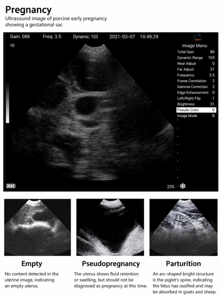
How to Recognize Pregnancy on the Screen
Interpreting the image correctly is the skill that separates an effective pregnancy check from an inconclusive one. Here is what to look for at each stage, based on published veterinary ultrasound literature.
28–35 days (cattle): The earliest reliable sign is the gestational sac — a dark, fluid-filled circle approximately 10–15 mm in diameter. It appears black because fluid does not reflect sound waves. Inside the sac, a small bright spot — the embryo — may be visible. By approximately day 30, a flickering bright dot at the center of the embryo indicates the fetal heartbeat. According to veterinary ultrasound reference texts, the gestational sac itself is typically first detectable between 26 and 28 days, with the embryo visible within the sac by approximately day 28–30 (Source:Youngquist & Threlfall, 2007, Current Therapy in Large Animal Theriogenology 2, pp. 294–302).
35–45 days (cattle): The embryo becomes more distinct. The amniotic membrane appears as a thin bright line surrounding the fetus. The placentomes — attachment points between the uterine wall and the placenta — begin to appear as C-shaped or oval bright structures, becoming visible in most pregnancies by day 35–40. These structures continue to enlarge and are readily identifiable by day 45 (Source:Fricke, 2002, Veterinary Clinics of North America: Food Animal Practice, 18(2): 233–254).
45–60 days (cattle): The fetus is clearly visible with recognizable head, body, and limb buds. Placentomes are larger and more numerous. The umbilical cord may be visible as a bright, coiled line. By this stage, fetal movement may be observed during real-time scanning.
Beyond 60 days (cattle): The fetus fills more of the screen. Skeletal structures like the skull, ribs, and spine appear bright white. At this stage, pregnancy is unmistakable even to a novice operator — but the economic value of early detection has already passed.
Key signs in sheep and goats (25–35 days): The gestational sac is visible transabdominally as a dark circular area. By 30–35 days, the embryo and heartbeat are detectable. Multiple gestational sacs indicate twins or triplets — important information for nutritional management of the ewe or doe. Portable ultrasound enables early pregnancy detection in sheep from approximately 25 days post-breeding, improving reproductive management in small ruminant systems (Source:Szenci et al., 2020, Animals, 10(7): 1164, PMC7363473).
What is NOT a pregnancy: A full bladder also appears as a dark fluid-filled area on the screen, which can be mistaken for a gestational sac by inexperienced operators. The key difference: a bladder is an irregular, pear-shaped structure, while a gestational sac is a clean circle with a defined wall. Proper operator training is essential; the 256-frame cine loop playback function allows frame-by-frame review of ambiguous images, reducing the risk of false-positive diagnoses from fluid-filled non-uterine structures (Fricke, 2002).
Pregnancy Detection Timeline Across Species
| Animal | Earliest Reliable Detection | Notes |
|---|---|---|
| Cattle | ~28 days | Gestational sac visible; heartbeat detectable from ~30 days |
| Sheep | ~25 days | Transabdominal; smaller probe may be needed for early scans |
| Goats | ~25 days | Similar to sheep; custom probe settings improve image quality |
| Swine | ~18 days | Earliest of livestock species; requires familiarity with sow anatomy |
| Horses | ~14–18 days | Rectal approach; early mobility examination possible |
Table 1: Earliest pregnancy detection timeframes by species using portable ultrasound
Common Questions
With a standard 3.5 MHz probe used rectally, pregnancy can be reliably detected as early as 28 days post-insemination in cattle. At this stage, the gestational sac is visible as a dark fluid-filled circle approximately 10–15 mm in diameter. Accuracy increases significantly after 35 days when the embryo and heartbeat are clearly identifiable. For sheep, detection is possible from approximately 25 days; for swine, from approximately 18 days.
Yes. Portable veterinary ultrasound scanners with IPX7-rated probes are designed for field use. An IPX7 rating means the probe can be submerged in water and cleaned with disinfectant solutions between animals without damage. The scanner body is sealed against moisture and dust. Battery life of 6+ hours supports a full day of scanning without access to power.
Yes. A portable livestock ultrasound scanner is suitable for multi-species use. The 3.5 MHz standard probe works well for cattle, horses, and larger livestock. For small ruminants like sheep and goats, the probe frequency can be customized for optimized near-field imaging. One device covers an entire mixed-species operation, from pregnancy checking cows to monitoring follicular activity in mares to measuring backfat in finishing lambs.
While formal training accelerates proficiency, the 256-frame cine loop playback feature significantly reduces the learning curve. A new operator can scan known-pregnant animals, replay the images frame by frame afterward, and learn what a positive pregnancy looks like at each gestational stage. Most AI technicians and veterinarians become confident in their readings after scanning 10–20 known-pregnant animals. Remote video guidance from an experienced practitioner can also assist with initial training.