Animal Type and Farm Routine Considerations
A cattle ultrasound machine is an important tool for reproductive management, pregnancy detection, and herd health evaluation on beef and dairy operations. However, the way you scan, when you scan, and what you look for can change depending on the type of animal and the daily farm routine. This article explains how factors like body size, behavior, feeding patterns, milk production schedules, and the intended use of the animal influence ultrasound practices. Whether you work with dairy cows, beef heifers, or breeding bulls, understanding these differences helps you get more reliable results from your ultrasound equipment.
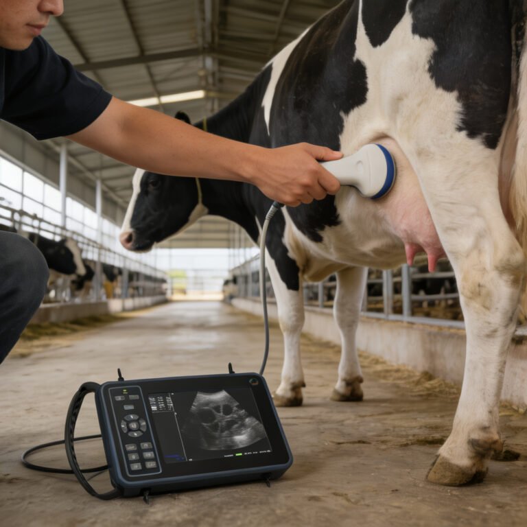
What a Cattle Ultrasound Machine Does on a Farm
A cattle ultrasound machine uses high-frequency sound waves to create images of internal structures, most commonly the reproductive tract. On a farm, it is used primarily for:
- Early pregnancy detection (as early as 26–30 days post-breeding).
- Fetal aging and viability checks.
- Identification of twins or reproductive abnormalities.
- Ovarian structure assessment (follicles, corpora lutea) for breeding management.
- Bull breeding soundness evaluation (accessory sex glands, testicular measurements).
According to the textbook Bovine Reproduction (2nd Edition), ultrasound has become a cornerstone of modern herd fertility programs, but the accuracy of findings depends heavily on operator skill and how well the procedure is adapted to the individual animal and production system.
How Animal Body Size Affects Ultrasound Scanning
Body size is one of the first variables a technician considers. Larger frame sizes and heavier body condition can make it harder to obtain a clear image.
- Mature beef cows often carry more backfat and a fuller rumen, which can push the reproductive tract forward, requiring a longer probe reach and more pressure to get good contact.
- Dairy cows, especially high-producing Holsteins, are generally leaner and may be easier to scan transrectally, but their size still requires proper restraint and a methodical approach.
- Heifers have smaller pelvic dimensions and less abdominal fill, making early pregnancy checks easier, but the rectum is narrower, so using a smaller probe or gentler technique is important to avoid injury.
- Bulls present a different challenge: ultrasound is used externally (transcutaneous) over the scrotum and internally for accessory glands. The size and thickness of the scrotal neck and body condition can affect image quality.
A common rule: when body condition score exceeds 3.5 on a 5-point scale, additional care is needed to differentiate between bowel gas, fat layers, and the uterus.
Animal Behavior and Handling Considerations
Cattle temperament directly impacts the safety of both the operator and the animal, as well as image quality. Agitated or stressed animals tense their abdominal muscles, which can displace the reproductive tract and make transrectal imaging more difficult.
What Varies by Animal Type
- Dairy cows accustomed to daily handling in the parlor may remain calm during scanning, especially if the procedure is performed in a familiar setting.
- Beef cows on extensive pasture systems may be less accustomed to close human contact, increasing the need for a good squeeze chute and quiet handling techniques.
- Heifers are often more nervous than mature cows; taking time to let them settle in the chute before inserting the probe reduces risk of rectal tearing.
- Bulls can be unpredictable. Some are docile, but others may react aggressively. Adequate head restraint and a sturdy chute are non-negotiable.
Low-stress handling principles, as outlined by many Extension livestock programs, recommend minimizing noise, using slow movements, and avoiding electric prods during ultrasound sessions. This helps keep the animal relaxed and improves image clarity.
Feeding and Drinking Patterns That Influence Scanning
What and when an animal eats or drinks can affect the quality of an ultrasound exam. A full digestive tract can push the uterus into the pelvic cavity or make it harder to locate.
Practical Guidelines
- Feed timing: For non-pregnant cows, scanning before the morning feeding when the rumen is less full often yields better images. In pregnant animals later in gestation, the gravid uterus is more cranial and may be partially obscured by a full rumen, so scheduling scans before large meals helps.
- Water intake: A very full bladder can displace the reproductive tract and mimic early pregnancy. Conversely, an empty bladder may make it harder to define the bladder-uterus interface in early pregnancy checks. Ideally, cattle should have access to water but not be overhydrated immediately before the scan.
- Milk production effects: Lactating dairy cows consume large volumes of water and feed, so their rumen fill is consistently higher. This may require more probe maneuvering and can slightly reduce early pregnancy detection rates compared to heifers.
For routine herd pregnancy checks on dairy farms, many veterinarians simply work through the herd as cattle come from the milking parlor, accepting that moderate rumen fill is normal and adjusting technique accordingly.
Impact of Milk Production Routine on Ultrasound Scheduling
Dairy farm routines revolve around milking schedules, and this affects when ultrasound examinations can be practically and efficiently performed.
Key Points for Dairy Operations
- Scheduling around milking: Cows are often scanned immediately after milking when they are already in the holding area or returning from the parlor. This reduces extra handling and stress. However, full udders before milking can make the cow uncomfortable, so scanning pre-milking is less common.
- Time constraints: In large dairies, the speed of scanning matters. Operators must be efficient, often using portable units with quick boot-up, to examine many cows in a short window between milkings.
- Stress and cortisol: Adding ultrasound scanning before milking could increase stress, potentially affecting let-down. Keeping the scanning area separate from the parlor but using a familiar route can help.
- Fresh cows vs. mid-lactation: Early lactation cows (first 30 days) may have a more difficult-to-image uterus due to involution and fluid. Timing the first post-calving reproductive scan requires waiting until uterine involution is advanced enough, typically around 25–30 days post-calving.
Beef Cattle vs. Dairy Cattle: Key Differences in Ultrasound Use
The table below compares how a cattle ultrasound machine is typically used in beef and dairy production systems. These differences stem from management goals, animal handling frequency, and breeding methods.
| Factor | Beef Cattle | Dairy Cattle |
|---|---|---|
| Primary goal | Pregnancy detection, fetal aging, heifer selection, carcass traits | Pregnancy detection, ovarian monitoring, early embryo loss detection |
| Scanning frequency | Often seasonal (after breeding season) | Weekly or bi-weekly herd checks |
| Handling environment | Pasture-based, portable chute, less frequent handling | Confined, parlor-based, accustomed to daily routine |
| Operator | Often a veterinarian or trained technician, sometimes the producer | Typically a veterinarian or specialized herd health technician |
| Body condition | Variable; higher backfat can hinder transrectal access | Generally leaner, easier rectal access |
| Timing of scan | Usually 30–90 days post-breeding | As early as 26–30 days post-insemination |
Heifers, Cows, and Bulls: What Changes for Each Animal Type
Different classes of cattle require adjustments in technique, probe selection, and even the purpose of ultrasound.
- Heifers (nulliparous): Smaller reproductive tract, less abdominal fill. Early pregnancy detection is easier, but care must be taken not to traumatize the rectum. Ovarian scanning is useful for pre-breeding evaluation.
- Multiparous cows: Larger uterus, especially in later gestation. A sector or convex probe with deeper penetration may be needed. Identification of twins is more critical because of higher twinning rates in certain breeds and nutritional management.
- Bulls: Ultrasound is used for breeding soundness exams. Transrectal scanning evaluates seminal vesicles, ampullae, and prostate. Transcutaneous scanning over the scrotum measures testicular diameter and detects abnormalities. Behavior is a major safety concern.
In all cases, using the right probe frequency (e.g., 5–7.5 MHz for transrectal, 7.5–10 MHz for bull reproductive tract) is essential. Lower frequencies penetrate deeper but offer less detail; higher frequencies give better resolution for surface structures.
Common Mistakes When Using a Cattle Ultrasound Machine
Avoid these errors to improve safety and accuracy:
- Skipping adequate fecal removal before transrectal scanning, which blocks sound transmission.
- Using excessive force or a dry probe, risking rectal perforation.
- Ignoring animal behavior; struggling cattle can injure themselves or the operator.
- Scanning at the wrong time: for early pregnancy, scanning before day 26 often yields false negatives.
- Misinterpreting a full bladder as an early gestational sac.
- Not adjusting machine settings (gain, depth, frequency) for the specific animal type.
- Failing to maintain the equipment: a damaged probe or dead battery can disrupt a planned herd check.
Final Takeaway
A cattle ultrasound machine is a versatile tool, but it does not work the same way for every animal. Body size, behavior, feeding patterns, and the daily rhythm of the farm all influence how you scan and what you see. Beef and dairy operations have different goals and schedules, and heifers, cows, and bulls each present unique anatomical and behavioral challenges. By adapting your technique to the animal type and farm routine, you can get clearer images, more accurate diagnoses, and better reproductive outcomes for the herd.
Frequently Asked Questions
Yes. While basic operation can be learned, accurate interpretation of reproductive images requires hands-on training. Many veterinarians and universities offer courses in bovine ultrasound techniques. Understanding anatomy, image artifacts, and pregnancy stages is essential to avoid misdiagnosis.
A 5–7.5 MHz linear rectal probe works for most transrectal exams in cows and heifers. For bulls, a higher frequency (7.5–10 MHz) microconvex or linear probe may be recommended for scrotal imaging. Probe selection depends on animal size, target depth, and desired detail.
For best image quality, scan before the morning feeding when the rumen is less full. In dairy herds, scanning after milking is convenient and reduces stress. Avoid scanning when cattle are overly hungry or thirsty, as agitation can reduce image clarity.
Most modern machines can detect pregnancy as early as 26–30 days post-breeding. However, false negatives are more common before day 30, so many producers choose to recheck later or wait until at least 35 days for higher accuracy.
When performed correctly, transrectal ultrasound is safe. Risks include rectal tearing if the probe is forced or the animal struggles. Proper restraint, adequate lubrication, and slow, gentle manipulation reduce risk. Operators should be trained in bovine anatomy and low-stress handling.
Yes, ultrasound is very effective at identifying twin pregnancies, often by visualizing two embryos or separate fetal heartbeats. Early twin detection allows for nutritional adjustments, as twin pregnancies require more careful management near calving.
On many dairies, cows are scanned after milking when they are already moving through handling areas. This minimizes extra labor and stress. Scanning before milking is less common because a full udder can make cows uncomfortable and may increase restlessness.
References
- Penn State Extension guide to Predicting Pregnancy Loss
- Penn State Extension guide to Minimizing Interservice Intervals
- MSD Veterinary Manual article on Pregnancy Determination in Goats
- MSD Veterinary Manual article on Management of Llamas and Alpacas
Related Guides in This Category
- What Is Vet Ultrasound and When Does It Make Sense on a Farm?
- What Is Portable Ultrasound Machine for Animals and When Does It Make Sense on a Farm?
- Equine Ultrasound Machine: What Changes by Animal Type and Farm Routine?
- Ultrasound Machine for Dogs: What Changes by Animal Type and Farm Routine?
- Cattle Ultrasound: What Changes by Animal Type and Farm Routine?
- What Is Portable Ultrasound Machine for Pregnancy and When Does It Make Sense on a Farm?
- Bovine Ultrasound: What Changes by Animal Type and Farm Routine?
- What Is Livestock Ultrasound Machine and When Does It Make Sense on a Farm?
- Ultrasound Machine for Cattle: What Changes by Animal Type and Farm Routine?
- What Is Ultrasound Machine for Animals and When Does It Make Sense on a Farm?