Key Concept and Practical Farm Use
A livestock ultrasound machine is a piece of equipment that uses high-frequency sound waves to see inside an animal’s body. On a farm, it is most often used for pregnancy checking, reproductive health assessment, and basic herd management decisions. For many livestock producers, it replaces guesswork with a real picture of what is happening inside their cows, ewes, or does.
This article explains what a livestock ultrasound machine actually is, how it differs from other types of ultrasound, and when it becomes a practical tool for a farm rather than an expensive gadget. It separates common confusions, gives a straight comparison with other pregnancy detection methods, and points toward the types and comparisons that deserve their own in‑depth guides.
What Is a Livestock Ultrasound Machine in Simple Terms?
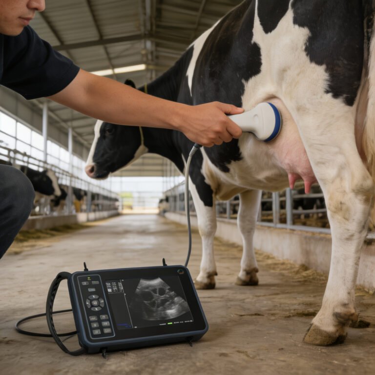
A livestock ultrasound machine is a portable imaging device built to work in a barn, pen, or pasture, not just in a veterinary clinic. It consists of a main unit (often a rugged tablet or console), a handheld probe (transducer), and a battery. The probe sends harmless sound waves into the animal and picks up the echoes that bounce back from different tissues. Those echoes are turned into a real‑time image on the screen.
In livestock, the most common probe type is the linear rectal transducer because it allows scanning through the rectal wall directly over the reproductive tract. According to the textbook Veterinary Ultrasound (Nyland & Nyland, 2nd Edition, Chapter 12), the linear‑array transducer is widely used for bovine reproductive scanning because of its near‑field resolution and wide field of view.
Farm ultrasound machines are not the same as human ultrasound equipment. They are designed to be dust‑resistant, shock‑resistant, and operable with gloves on. They also run on rechargeable batteries because many cattle sheds and lambing pens have no convenient power outlet nearby.
How Does a Livestock Ultrasound Machine Work?
Ultrasound works like sonar. The transducer emits short bursts of sound at a frequency far above human hearing. When those sound waves hit tissue boundaries – fluid, muscle, fat, bone, an early foetus – they reflect back. The machine measures the time it takes for each echo to return and builds a two‑dimensional grey‑scale image.
On a farm, the most common target is the pregnant uterus. Fluid appears black, tissue appears grey, and bone or dense structures appear white. An experienced operator can see the amniotic fluid, foetal shape, and even a heartbeat after a certain number of days.
No radiation is involved, and the procedure is considered safe for both the animal and the operator when proper restraint and hygiene are used.
What Common Confusions Should Farmers Avoid?
Several misunderstandings can lead to unrealistic expectations or poor equipment choices. The most common ones are:
- Ultrasound is not an X‑ray. It does not show bone in detail or replace a radiograph. It excels at soft‑tissue imaging, which is exactly what is needed for pregnancy diagnosis.
- A human ultrasound machine will not work well on a farm. Clinic‑grade cart‑based machines are not built for dirt, moisture, or constant movement. Farm ultrasound needs to be portable and rugged.
- Not every probe fits every animal. The probe shape, frequency, and scanning depth must match the species and the job. A probe made for small‑animal abdominal scanning is not suitable for rectal pregnancy checking in beef cows.
- Ultrasound does not replace good handling. Even the best image is useless if the animal is stressed or the operator cannot safely position the probe.
- Scanning livestock is not the same as scanning a dog or cat. The anatomy, restraint needs, and image interpretation are different, which is why farm‑specific training is important.
When Does a Livestock Ultrasound Machine Make Sense on a Farm?
A livestock ultrasound machine becomes a sensible tool whenever the value of knowing an animal’s reproductive status outweighs the cost and time of learning to operate it. Concrete farm scenarios where ultrasound adds real value include:
- Early, accurate pregnancy detection. Skilled operators can detect pregnancy in cattle as early as 28–30 days, weeks ahead of a blood test or manual palpation. This lets a farm cull open animals sooner, saving winter feed and labour.
- Identifying twin pregnancies. Ultrasound can spot twins in sheep or cattle, which changes nutrition and management before lambing or calving.
- Checking foetal viability and age. Seeing a heartbeat and measuring foetal size helps confirm a viable pregnancy and estimate the calving or lambing window.
- Reproductive tract scoring. In heifer selection, ultrasound can evaluate ovarian structures and uterine health, supporting better breeding decisions.
- Bull breeding soundness support. While not a replacement for a full BSE, ultrasound can assess testicular structure and help identify abnormalities.
- Managing a large herd or flock efficiently. Farms that run hundreds of animals often invest in their own machine to avoid the per‑head cost and scheduling delays of a contracted scanner.
- Improving biosecurity and data control. On‑farm scanning means no outside vehicle and no sharing of digital records with a third party.
For a small hobby farm with only a few animals, paying for occasional professional scanning may remain the practical choice. But as herd size grows or reproductive performance becomes a profit driver, owning a livestock ultrasound machine starts to look like precision management rather than a luxury.
Livestock Ultrasound vs. Other Pregnancy Detection Methods
No single method fits every farm. The table below compares the four most common ways to determine pregnancy in cattle and small ruminants, so you can see where ultrasound sits in the decision process.
| Method | Earliest Reliable Detection (Cattle) | Accuracy | Equipment Need | Skill Level Required | Animal Stress |
|---|---|---|---|---|---|
| Ultrasound (rectal, B‑mode) | 28–30 days | Very high (operator dependent) | Ultrasound machine and probe | Moderate to high; needs training | Moderate (requires restraint and rectal entry) |
| Manual rectal palpation | 35–45 days | High in experienced hands | Glove and lubricant only | High; takes many months to master | Moderate |
| Blood pregnancy test (PAG‑based) | 28–30 days | Very high (lab accuracy) | Blood collection supplies + lab service | Low; sample is easy to collect | Low |
| Observation of return to oestrus | 21+ days after breeding | Moderate to low; many false negatives | None | High observational skill | None |
Ultrasound stands out because it gives immediate results and provides visual information beyond a simple yes‑or‑no pregnancy answer. That visual data often justifies the higher upfront equipment investment for farms where reproductive efficiency is a key performance indicator.
Key Features to Understand Before Looking at Machine Types
Before diving into a comparison of specific ultrasound machines, it helps to understand the basic features that separate one livestock ultrasound machine from another. These are the aspects that will show up again when you later read about “types of livestock ultrasound machines” and “how to choose a probe.”
- Transducer (probe) shape and frequency. Linear rectal probes are the workhorse for cattle. Convex probes are often used for trans‑abdominal scanning in sheep and goats. Frequency (MHz) affects how deep the sound penetrates and how much detail you see. Higher frequency sees more detail but less depth; lower frequency goes deeper but with less resolution.
- Portability and battery life. Most farm machines are handheld or tablet‑based. Check how long the battery lasts during continuous scanning, because a morning of pregnancy checking can drain a weak battery fast.
- Screen size and outdoor visibility. Sunlight washes out many screens. A large, bright display with anti‑glare coating matters when scanning in an open‑front barn or pasture.
- Durability and waterproof rating. Look for dust‑proof and water‑resistant seals. Farm conditions are tough on electronics.
- Image storage and connectivity. Machines that let you save images and transfer them to a computer or phone make record‑keeping simpler and allow a second opinion from a veterinarian.
All of these features will be expanded in a dedicated article that compares specific livestock ultrasound machine types and their best‑fit farm situations. This overview simply gives you the vocabulary to start asking the right questions.
Common Mistakes and Farm Realities
Even a good livestock ultrasound machine can fail to deliver value if it is used without a clear plan. These are the pitfalls that trips up the most first‑time users.
- Buying a machine before securing training. The image on the screen means nothing if you cannot tell a pregnancy from a full bladder. Most university Extension programs and veterinary practices offer ultrasound training courses.
- Using the wrong probe for the species. A linear rectal probe made for cows will not fit safely or image correctly in a ewe. Matching probe to species is non‑negotiable.
- Skipping animal restraint. Cattle need to be caught in a crush or head‑gate. Trying to scan a loose cow in a pen is dangerous for the operator and the animal, and the image will be unreadable.
- Relying on ultrasound alone for all reproductive calls. Ultrasound is a tool, not a crystal ball. It works best inside a broader reproductive management programme that includes body condition scoring, good nutrition, and proper bull or AI management.
- Ignoring hygiene and biosecurity. Rectal probes must be cleaned and disinfected between animals. Dirty probes spread uterine infections, which is exactly the opposite of what any breeding herd needs.
Where to Go Next: Types, Probes, and Comparisons
This article set out to answer the two core questions: “What is a livestock ultrasound machine?” and “When does it make sense on a farm?” If you now have a clearer picture of what the equipment does and whether it fits your operation, the next natural step is to look at the different types of machines, probe options, and how they compare for specific livestock species and farm sizes.
Those deeper comparisons are handled in separate guides that look at portable versus cart‑based ultrasound, linear versus convex probes, and the practical differences between scanning cattle, sheep, goats, and swine. For now, the main takeaway is that a livestock ultrasound machine is not just a device; it is an entry point to better reproductive data and smarter farm decisions when matched to the right training and the right farm context.
Frequently Asked Questions
Yes. Even a simple portable machine requires proper training to interpret images accurately. Many university Extension programmes, veterinary colleges, and experienced livestock scanning services offer hands‑on courses.
No, diagnostic ultrasound uses harmless sound waves, not radiation. The procedure is safe when proper restraint and clean techniques are used. Stress from poor handling poses a greater risk than the ultrasound itself.
A farm ultrasound is built for portable, outdoor use: it is battery‑powered, rugged, and often comes with probes that work through the rectal wall or abdominal skin of large animals. A clinic ultrasound is usually cart‑based, less mobile, and designed for a clean, controlled environment.
For cattle, a linear rectal probe is standard because it provides the near‑field resolution needed when scanning the reproductive tract through the rectal wall. For sheep and goats, a convex abdominal probe is more common because the reproductive tract is scanned trans‑abdominally. Probe frequency and probe shape must match the animal’s size and the entry route.
With a good machine and a skilled operator, pregnancy can be detected as early as 28–30 days after breeding in cattle. Foetal viability and heartbeat become clearly visible around day 35–40. Early detection allows farmers to make timely culling and feeding decisions.
Very important. Most livestock ultrasound work happens in pens, handling facilities, or chutes where access to mains power is limited. A lightweight machine with a long‑lasting battery lets you scan a whole group of animals without stopping to recharge.
The most common mistake is attempting to use the machine without any hands‑on training. Misreading an image can lead to calling an open cow pregnant and feeding it for months with no calf produced, which directly hurts farm profitability.
References
- Penn State Extension guide to Predicting Pregnancy Loss
- Penn State Extension guide to Minimizing Interservice Intervals
- MSD Veterinary Manual article on Pregnancy Determination in Goats
- MSD Veterinary Manual article on Management of Llamas and Alpacas
Related Guides in This Category
- Ultrasound Machine for Cattle: What Changes by Animal Type and Farm Routine?
- Equine Ultrasound Machine: What Changes by Animal Type and Farm Routine?
- What Is Portable Ultrasound Machine for Animals and When Does It Make Sense on a Farm?
- What Is Ultrasound Machine for Animals and When Does It Make Sense on a Farm?
- What Is Portable Ultrasound Machine for Pregnancy and When Does It Make Sense on a Farm?
- Bovine Ultrasound: What Changes by Animal Type and Farm Routine?
- Cattle Ultrasound Machine: What Changes by Animal Type and Farm Routine?
- Sheep Ultrasound Machine: What Changes by Animal Type and Farm Routine?
- What Is Portable Ultrasound Machine Veterinary and When Does It Make Sense on a Farm?
- What Is Vet Ultrasound and When Does It Make Sense on a Farm?