Key Concept and Practical Farm Use
For many farmers and ranchers, the term “vet ultrasound” simply means using sound‑wave technology to see inside an animal without surgery. On a farm, the most common reason is pregnancy detection, but the tool is also used for reproductive health checks, monitoring fetal development, and diagnosing certain internal conditions. This article explains what vet ultrasound is in plain farm language, when it actually makes sense to call a veterinarian with an ultrasound scanner, and what to expect from the procedure. It is written for cattle, sheep, goat, pig, and horse owners who want practical reproductive management information—not a sales pitch for equipment.
What Does “Vet Ultrasound” Actually Mean on a Farm?
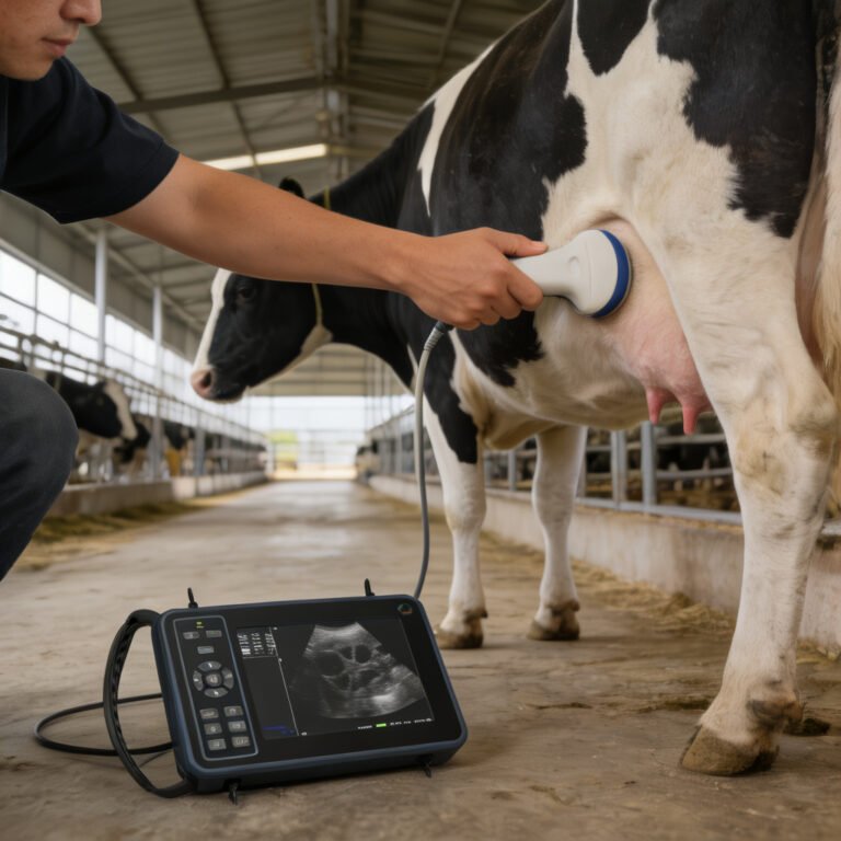
Veterinary ultrasound uses high‑frequency sound waves to create real‑time images of soft tissue inside an animal’s body. A handheld probe (transducer) is placed against the skin or inserted rectally; it sends out sound waves that bounce off internal structures, and the echoes are converted into an image on a screen. No radiation is involved, and the exam is considered non‑invasive.
On a farm, the focus is usually on the reproductive tract—confirming pregnancy, checking fetal number and viability, evaluating ovarian structures, or identifying uterine infections. While the technology works the same way as in human medicine, farm‑oriented ultrasound machines are often designed to be portable, battery‑powered, and rugged enough for a barn or chute‑side environment. According to the textbook Reproduction in Farm Animals (7th Edition, Chapter 14), real‑time B‑mode ultrasound has become the standard tool for early pregnancy diagnosis in cattle, sheep, goats, and pigs.
When Does a Vet Ultrasound Exam Make Sense?
Not every animal needs an ultrasound, and not every farm visit requires one. The exam makes the most sense when precise, early information changes a management decision. Common on‑farm scenarios include:
- Early pregnancy detection—Confirming pregnancy earlier than rectal palpation or blood tests allows, which can shorten re‑breeding intervals and identify open animals sooner.
- Fetal viability checks—Monitoring heartbeat, fetal movement, and placental health, especially in high‑value animals or after a stressful event.
- Twin detection—In cattle or small ruminants, identifying twins early helps adjust nutrition and plan for assisted births.
- Reproductive tract evaluation—Assessing ovarian status, diagnosing cystic ovaries, endometritis, or other issues that affect fertility.
- Targeted health checks—In some cases, ultrasound can visualize abscesses, fluid accumulation, or conditions like hardware disease when the probe is placed on the abdominal wall.
If knowing whether an animal is pregnant, how many fetuses she carries, or the reason she is not cycling will influence culling, feeding, or marketing decisions, then a vet ultrasound exam often pays for itself in better management outcomes.
Vet Ultrasound vs. Other Farm Pregnancy Detection Methods
Farmers often choose between several pregnancy detection approaches. The table below compares the most common ones with ultrasound.
| Method | How It Works | Earliest Reliable Stage | Main Advantages | Main Limitations | Farmer Takeaway |
|---|---|---|---|---|---|
| Vet Ultrasound | Sound‑wave imaging of uterus and fetus | Cattle: ~28‑35 days (typical for experienced operator); small ruminants: ~25‑30 days | Visual confirmation of fetus, heartbeat, twins; immediate result; non‑invasive | Requires skilled operator and appropriate equipment; can be more expensive per head than palpation | Best when early, precise reproductive information is needed |
| Rectal Palpation | Manual feeling of reproductive tract through rectal wall | Cattle: ~35‑45 days (depending on operator) | Low equipment cost, widely available, fast for large herds | Later detection window, limited fetal viability info, operator fatigue, small risk of injury | Solid, cost‑effective choice for routine herd checks in cattle when early detection isn’t critical |
| Blood Tests (e.g., pregnancy‑specific protein B or PSPB) | Lab analysis of blood sample for pregnancy markers | Cattle: ~28‑30 days; sheep/goats: ~30‑35 days | No need to restrain for exam; samples can be shipped; useful for extensive systems | Delayed results, cannot determine fetal viability or twins, false positives possible after pregnancy loss | Good when handling is difficult or ultrasound access is limited |
| Behavioral Observation (non‑return to estrus, visual changes) | Watching for heat cycles, udder development, abdominal shape | Not reliable for early detection; varies by species | No equipment or handling needed | Very imprecise, late detection, cannot confirm pregnancy itself | Only a clue, not a diagnostic method; should not be used alone for management decisions |
In practice, many farms combine methods: ultrasound for individual high‑value animals or problem cases, and palpation or blood tests for routine herd‑level management.
Common Types of Vet Ultrasound Scanners Used on Farms
Vet ultrasound machines come in many forms, but on a farm you will typically see portable or handheld units designed for field work. Without going into product detail, a few broad categories help explain what a farmer might encounter:
- Linear‑array rectal probes—Commonly used for transrectal pregnancy diagnosis in cattle and horses. They produce high‑resolution images of superficial structures like the early pregnancy sac.
- Convex (sector) probes—Often used transabdominally in small ruminants, pigs, and sometimes cattle. They offer a wider field of view for scanning deeper structures.
- Handheld all‑in‑one units—Battery‑operated, compact scanners that combine screen and probe, designed for chute‑side use.
The critical factor is not the machine’s brand but whether the probe type and frequency match the species and the gestational stage. A separate guide can explore different ultrasound scanner types in greater depth for those who need to compare specifications.
What a Farmer Should Expect During a Farm Ultrasound Visit
When a veterinarian brings an ultrasound scanner to the farm, the priority is animal restraint and safety. For cattle and horses, the exam is usually transrectal, meaning the vet inserts a lubricated probe into the rectum to image the reproductive tract. For small ruminants and pigs, a transabdominal approach (placing the probe on the clipped, gel‑coated skin of the lower flank or abdomen) is more common.
A typical visit follows these steps:
- Restraint—The animal is securely held in a chute, head gate, or handling crate. Calm animals give better images.
- Preparation—For abdominal scans, hair may be clipped and ultrasound gel applied. For rectal scans, fecal material is gently evacuated.
- Scanning—The vet moves the probe to locate the uterus, fetus, fluid, or heart motion. The whole process usually takes 1–5 minutes per animal.
- Immediate result—Pregnancy status, fetal viability, and sometimes fetal sex (in experienced hands) are visible on the screen. The farmer sees the image in real time.
The key is good animal handling. A stressed or moving animal makes it harder to get a clear image and increases the risk of injury for both the animal and the handler.
Key Factors That Affect Ultrasound Usefulness on a Farm
Ultrasound is not a magic window. Its value depends on several practical factors:
- Gestational stage—Too early (before around 25 days in cattle) may give a false negative; after mid‑pregnancy the fetus drops over the pelvic brim, making transrectal scanning harder.
- Animal size and body condition—Overly fat animals can scatter the sound beam, reducing image quality.
- Operator skill—An experienced veterinarian or technician can reliably detect pregnancy early; a novice operator may miss structures.
- Equipment capability—Machines with appropriate probe frequencies and penetration depth are needed for different species and gestational stages.
- Animal cooperation—Restless or aggressive animals lead to poor images and potential safety incidents.
- Facilities—A well‑designed handling system with good lighting and non‑slip footing makes the examination smoother.
Understanding these limitations helps a farmer decide when an ultrasound exam is likely to yield reliable information and when another method might be more practical.
Does Every Farm Need Vet Ultrasound?
No. Vet ultrasound is a management tool, not a universal requirement. A small homestead with a few animals may only need a veterinarian to confirm pregnancy once a year, and a blood test or palpation might suffice for that. On the other hand, a commercial beef or dairy operation that tightly manages breeding windows, culling, and nutrition often benefits from routine ultrasound scanning because early pregnancy information directly impacts profitability.
The decision to use ultrasound should be based on:
- How much a missed or late pregnancy costs the operation.
- The value of knowing fetal number or ovarian status.
- Whether early open detection allows timely re‑breeding.
- The availability of skilled ultrasound veterinarians in the area.
If the cost of the exam is less than the cost of keeping an open cow for an extra season, the economic case is strong. Many extension services suggest evaluating the payback period: if ultrasound information prevents the expense of feeding a non‑productive animal, it rapidly covers its own cost.
Common Mistakes When Considering Farm Ultrasound
Farmers sometimes expect ultrasound to solve every reproductive mystery, but a few common missteps can undermine its effectiveness:
- Scanning at the wrong time—Checking too early (before the conceptus is visible) or too late (when the fetus is out of range) leads to inconclusive or false results.
- Assuming a negative ultrasound means the animal is permanently infertile—A single negative scan on a given day does not mean the animal cannot conceive; it might just be at the wrong stage of the cycle.
- Using poor restraint—An animal that kicks or struggles not only risks injury but also degrades image quality, leading to misdiagnosis.
- Expecting a product‑like “yes/no” pregnancy test —Ultrasound provides visual evidence, but interpreting early pregnancy requires skill; sometimes ambiguous images occur.
- Relying on ultrasound alone to fix a herd fertility problem—Ultrasound diagnosis should be part of a broader reproductive management plan that includes nutrition, breeding soundness exams, and herd health protocols.
Being realistic about what the technology can and cannot do sets the stage for a productive veterinary visit.
Vet Ultrasound in Different Livestock Species
Although the principles are the same, practical ultrasound differs slightly among species:
- Cattle—Transrectal scanning is standard. Pregnancy can be reliably confirmed from around 28 days. Fetal sexing is possible around 55–70 days by an experienced operator.
- Sheep and goats—Transabdominal scanning is typical, often with the animal standing or in a sitting position. Early diagnosis at 25–30 days is common. Real‑time scanning enables better flock management.
- Pigs—Transabdominal ultrasound is used, usually from 21–28 days post‑breeding. The procedure is quick, but individual pig handling can be a challenge.
- Horses—Transrectal ultrasound is the gold standard for early pregnancy and twin detection from about 14–16 days. Twin reduction is possible if detected early.
The key takeaway is that the approach is tailored to each species’ anatomy and typical management style. A veterinarian with species‑specific ultrasound experience is essential.
Final Takeaway
Vet ultrasound is a valuable diagnostic tool that gives farmers a real‑time look inside their animals’ reproductive tracts. It makes the most sense when early, accurate pregnancy information or reproductive health insights will directly change a management decision—be it culling an open cow, adjusting nutrition for a pregnant ewe, or scheduling a re‑check for a mare. However, ultrasound is not a substitute for overall herd health planning, good handling facilities, or skilled veterinary support. Used at the right time and for the right reasons, it can be a powerful aid in profitable and efficient livestock farming.
References
- Penn State Extension guide to Predicting Pregnancy Loss
- Penn State Extension guide to Minimizing Interservice Intervals
- MSD Veterinary Manual article on Pregnancy Determination in Goats
- MSD Veterinary Manual article on Management of Llamas and Alpacas
Related Guides in This Category
- What Is Portable Ultrasound Machine for Pregnancy and When Does It Make Sense on a Farm?
- Ultrasound Machine for Dogs: What Changes by Animal Type and Farm Routine?
- What Is Portable Ultrasound Machine for Animals and When Does It Make Sense on a Farm?
- What Is Portable Ultrasound Machine Veterinary and When Does It Make Sense on a Farm?
- Bovine Ultrasound: What Changes by Animal Type and Farm Routine?
- What Is Livestock Ultrasound Machine and When Does It Make Sense on a Farm?
- Cattle Ultrasound Machine: What Changes by Animal Type and Farm Routine?
- Cattle Ultrasound: What Changes by Animal Type and Farm Routine?
- What Is Ultrasound Machine for Animals and When Does It Make Sense on a Farm?
- Equine Ultrasound Machine: What Changes by Animal Type and Farm Routine?