Animal Type and Farm Routine Considerations
Cattle ultrasound is a tool for pregnancy detection, reproductive assessment, and herd management. But using it effectively depends on more than just the machine. Animal body size, behavior, feeding patterns, and daily farm routines all change how you scan and what you see. A dairy cow in a milking parlour isn’t the same as a beef cow in a pasture. A heifer can be harder to scan than a mature cow. And a full rumen can interfere with image quality.
This article walks through what changes when you use cattle ultrasound across different animal types and farm routines. You’ll find practical comparisons, handling tips, and a checklist to help you plan better scanning sessions.
How Animal Body Size Affects Cattle Ultrasound
Body size influences probe frequency, scanning window, and image depth. Larger animals need lower frequencies to penetrate deeper tissue. Smaller animals, like calves or young heifers, often give clearer images with higher‑frequency linear probes.
General guidelines by animal size:
- Mature beef cows (over 600 kg): Use a 3.5 MHz sector or convex probe for deep uterine imaging.
- Mature dairy cows (500–700 kg): A 5.0 MHz linear rectal probe often works well because the reproductive tract sits relatively forward.
- Heifers (under 400 kg): A 5.0–7.5 MHz linear probe provides better detail. The reproductive tract is shallower, so high frequency gives clearer early pregnancy images.
- Calves or small animals: A linear 7.5 MHz probe is best for shallow scanning, but reproductive scanning in very young calves is uncommon outside research.
According to Large Animal Clinical Procedures for Veterinary Technicians (3rd Edition, Chapter 11), transducer selection must match the depth of the target structure. Using the wrong frequency is a common error that leads to missed diagnoses.
What Changes with Animal Behavior and Temperament?
Behavior is one of the biggest variables. Excitable or aggressive cattle make scanning dangerous and reduce image quality. Calm animals allow longer, more accurate scans.
Handling and restraint need to match the animal:
- Calm dairy cows used to daily handling may only need a headlock or a simple chute with a bar behind.
- Range beef cows may require a squeeze chute with side gates and a head catch for safety.
- Brahman‑influenced breeds tend to be more excitable and often need tighter restraint and a faster scanning approach.
- Heifers are often more nervous than older cows and benefit from a quiet working environment and minimal noise.
- Bulls used for breeding soundness exams need extra care. Use a sturdy chute that limits lateral movement.
The Beef Cattle Science handbook (9th Edition, Chapter 10) notes that low‑stress handling improves reproductive efficiency and reduces scanning artifacts caused by animal movement.
How Feeding and Drinking Patterns Influence Ultrasound Timing
What and when an animal eats or drinks changes what you see on ultrasound. A full rumen pushes the reproductive tract forward or sideways. A full bladder can obscure early pregnancy structures.
Practical timing tips:
- For pregnancy diagnosis, scan before morning feeding when the rumen is less distended.
- If you must scan after feeding, wait at least 2–3 hours.
- In dairy herds on total mixed rations, the rumen stays fuller longer. Consider scanning during the milking session when the rumen may be slightly emptier.
- Avoid scanning immediately after cattle drink large amounts of water; a full bladder can mimic a fluid‑filled structure.
According to Veterinary Reproduction and Obstetrics (10th Edition, Chapter 8), optimal scanning conditions include an empty rectum and minimal rumen fill to improve uterine visibility.
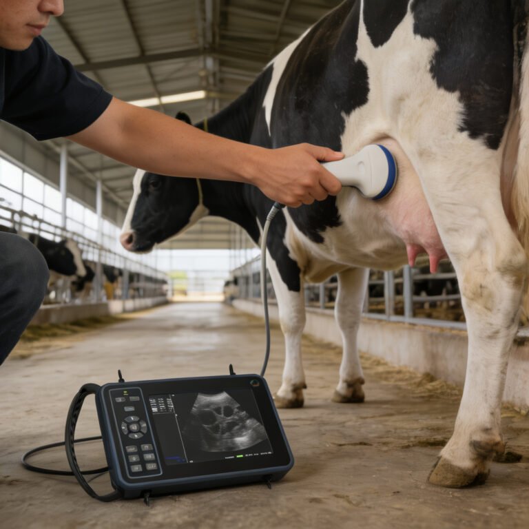
Ultrasound Adjustments for Milk Production Routine
Dairy cows present unique challenges. The udder is large, and in late gestation the gravid uterus can be tucked far forward. Milking schedule also affects handling and image quality.
Key adjustments for dairy ultrasound:
- Scan after milking whenever possible. An empty udder makes rectal palpation and probe placement easier.
- In early lactation, a full udder can press the uterus forward. Use a longer‑reach probe or reposition the cow to tilt the pelvis slightly.
- Dairy cows in tie‑stall barns can be scanned in place, but a head catch is still needed if the cow is restless.
- In high‑producing cows, the reproductive tract may be slower to involute post‑calving. Wait at least 30 days after calving for accurate pregnancy diagnosis.
Species‑Specific Scanning: How Cattle Compare to Other Livestock
Cattle ultrasound isn’t the same as scanning sheep, goats, or pigs. Each species has different anatomy, size, and handling requirements.
| Factor | Cattle | Sheep/Goats | Pigs |
|---|---|---|---|
| Probe placement | Transrectal (most common) | Transabdominal (external) | Transabdominal (external) |
| Frequency range | 3.5–7.5 MHz | 5.0–7.5 MHz | 3.5–5.0 MHz |
| Restraint needed | Chute or headlock | Manual hold or cradle | Snout snare or pen corner |
| Main use | Pregnancy, reproductive tract | Pregnancy, fetal counting | Pregnancy, backfat, loin depth |
| Handling difficulty | Moderate to high | Low to moderate | High (squealing, movement) |
For cattle, transrectal scanning is standard because the large body wall attenuates abdominal ultrasound. Sheep and goats are small enough for external scanning. Pigs are challenging due to thick fat layers and resistance to restraint.
Common Mistakes When Using Ultrasound on Cattle
Even with good equipment, mistakes happen. Knowing what to avoid can save time and reduce misdiagnosis.
- Wrong probe for the animal size. A 7.5 MHz probe on a large beef cow won’t penetrate deep enough.
- Scanning too early after breeding. Pregnancy is not reliably visible before 28 days (fetal heartbeat) with most portable scanners.
- Poor contact or lubrication. Dry rectal entry causes air gaps and poor image quality.
- Moving probe too fast. A slow, systematic sweep reduces missed findings.
- Ignoring animal stress. Struggling cattle give blurry images and can injure the operator or damage the probe.
- Not cleaning the probe properly between animals. This increases disease transmission risk.
Checklist for a Smooth Cattle Ultrasound Session
Use this checklist before you start scanning.
- Confirm handling facility is safe: chute, head catch, and rear gate working.
- Select probe based on animal size and target depth (3.5–7.5 MHz).
- Check ultrasound machine battery and cable connections.
- Have enough ultrasound gel or obstetric lubricant ready.
- Organize animal records and ear tags for easy recording.
- Plan animal flow: bring only a few animals at a time to keep them calm.
- Wear protective gloves and boots; consider a plastic sleeve for the probe handle.
- Have disinfectant and paper towels for probe cleaning between animals.
- For dairy, schedule scans after milking if possible.
- For breeding soundness, have a second person to assist with bull handling.
When these steps are followed, scanning time per cow often drops by 30% and image quality improves noticeably.
Frequently Asked Questions
Prices vary widely, but basic portable units suitable for cattle pregnancy detection start at a few thousand dollars. More advanced scanners with reproductive measurement tools cost more. Many farms share equipment with a local vet instead of buying their own.
It is possible with calm, well‑handled dairy cows in headlocks, but for safety, two people are better. One person restrains the cow while the other scans. In chutes, a rear bar helps but an assistant is still recommended.
Fetal heartbeat can be seen as early as 28 days post‑breeding with a skilled operator and a good‑quality 5.0–7.5 MHz linear probe. Most practical on‑farm scanning targets day 30–35 for reliable results.
Yes. Bos indicus breeds often have more intra‑abdominal fat and a tighter cervix, which may slightly reduce early pregnancy visibility. Thick‑muscled beef breeds may require lower frequencies than dairy breeds.
Clean the probe between every animal to prevent cross‑contamination. Use a disinfectant recommended by the manufacturer, rinse thoroughly, and dry the probe head before the next scan.
Yes, if the machine has interchangeable probes. Use a linear 5.0–7.5 MHz probe for sheep transabdominal scanning and switch to a 3.5–5.0 MHz sector or linear probe for cattle transrectal scanning. Always clean probes thoroughly between species.
Scanning too hastily. New operators often move the probe too quickly and miss the uterus or fetal structures. A slow, methodical sweep with good lubricant and proper probe placement is essential.
References
- Penn State Extension guide to Predicting Pregnancy Loss
- Penn State Extension guide to Minimizing Interservice Intervals
- MSD Veterinary Manual article on Pregnancy Determination in Goats
- MSD Veterinary Manual article on Management of Llamas and Alpacas
Related Guides in This Category
- Cattle Ultrasound Machine: What Changes by Animal Type and Farm Routine?
- Equine Ultrasound Machine: What Changes by Animal Type and Farm Routine?
- What Is Portable Ultrasound Machine for Animals and When Does It Make Sense on a Farm?
- What Is Ultrasound Machine for Animals and When Does It Make Sense on a Farm?
- What Is Livestock Ultrasound Machine and When Does It Make Sense on a Farm?
- Bovine Ultrasound: What Changes by Animal Type and Farm Routine?
- Sheep Ultrasound Machine: What Changes by Animal Type and Farm Routine?
- What Is Vet Ultrasound and When Does It Make Sense on a Farm?
- What Is Portable Ultrasound Machine for Pregnancy and When Does It Make Sense on a Farm?
- What Is Portable Ultrasound Machine Veterinary and When Does It Make Sense on a Farm?