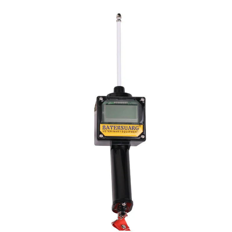
Key Concept and Practical Farm Use
A portable ultrasound machine veterinary is a compact, lightweight imaging tool designed for field use on farms. It lets you see inside an animal’s body—most often to check pregnancy, assess reproductive health, or evaluate internal structures—right where the animal stands, without moving her to a clinic.
For farm managers, herd owners, and large‑animal practitioners, the main question isn’t just “What is this machine?” but “When does it actually make sense to own or use one?” This article answers that plainly. You’ll get a straightforward definition, practical use cases, a comparison with cart‑based systems, what to look for, and common mistakes to avoid. We won’t sell you a machine or recommend specific models. Instead, you’ll learn how to decide if a portable ultrasound fits your regular farm routine.
What Is a Portable Ultrasound Machine Veterinary?
A portable ultrasound machine veterinary is a handheld or battery‑powered device that uses high‑frequency sound waves to create real‑time images of soft tissues, organs, and fluid‑filled structures inside an animal’s body. Unlike a cart‑based unit that stays in a clinic, a portable machine is built to travel. Most weigh under 5 kg (11 lb) and can be carried in a backpack or over the shoulder.
The main parts are:
- Console or tablet: the processing unit and screen for viewing images.
- Transducer (probe): the part you place against the animal. Different shapes and frequencies serve different purposes—linear probes for tendons and small parts, convex or sector probes for deep abdominal or reproductive scanning.
- Battery pack: often rechargeable, allowing several hours of cordless scanning.
- Software and storage: basic onboard measurement tools, video recording, and image saving for later review.
In livestock farming, these machines are most often used for pregnancy diagnosis, but they can also help detect uterine infections, monitor ovarian activity, or check for bladder stones and other internal issues. The images aren’t as detailed as a high‑end stationary system, but for routine field decisions, they provide enough information quickly.
If you are planning a purchase, you can see the full specifications of our cattle ultrasound machine.
When Does Portable Ultrasound Make Sense on a Farm?
You don’t need a portable ultrasound machine just because the technology exists. It makes sense when these four conditions overlap:
- You regularly need answers that change your management decisions. For example, confirming pregnancy early lets you sort open cows, change feeding groups, and plan rebreeding. If that information pays for itself quickly across your herd, the machine earns its place.
- Waiting for a veterinarian or travelling to a clinic costs more than the scan. In remote areas or during busy seasonal windows, having an operator on‑site can save days of delay and reduce animal stress.
- You have a trained operator or are willing to invest in training. Without basic ultrasound skills, the machine turns into an expensive paperweight. Interpretation errors can lead to expensive mistakes.
- Your workload justifies the battery and portability. If you’re scanning animals in pastures, handling facilities, or distant pens where power outlets don’t exist, a portable unit is the only practical option.
For a small herd with easy access to a large‑animal vet, a shared service arrangement or an occasional vet visit may make more sense than purchasing equipment. The decision isn’t about the machine’s features—it’s about whether the information changes your daily choices often enough.
Portable vs Cart‑Based Veterinary Ultrasound: A Quick Comparison
Not all “portable” machines are truly field‑ready, and many farmers confuse them with the stationary systems used in vet clinics. The table below highlights the practical differences that affect farm use.
| Factor | Portable Ultrasound Machine Veterinary | Cart‑Based Veterinary Ultrasound |
|---|---|---|
| Mobility | Handheld, battery‑operated, fits in a bag | Wheeled cart, tethered to power outlet |
| Weight | Typically 2‑5 kg (4‑11 lb) | Often 50‑100+ kg (110‑220 lb) |
| Image quality | Adequate for routine pregnancy, uterine health, basic abdominal scans | Higher resolution, more advanced image modes |
| Battery life | 2‑6 hours of continuous scanning | Mains power only |
| Best use | On‑farm pregnancy checks, field diagnostics, chute‑side work | Detailed exams, guided procedures, referral work |
| Main drawback | Limited depth or detail in very large or heavy‑conditioned animals | Impractical for farm calls, requires transport and setup |
For most livestock pregnancy work and routine field diagnostics, a portable ultrasound machine veterinary strikes the right balance between usability and image quality. Cart‑based units remain the gold standard for small‑animal clinics or specialized equine reproduction facilities, but they aren’t built for mud, dust, and tail‑gate operation.
Farm units vs clinic machines
Stationary ultrasound machines are what you often see in a vet clinic: a cart‑based system with a large monitor, multiple probes, and advanced imaging software. They are built for high‑resolution diagnostic work, but they are heavy, need wall power, and are typically too expensive and impractical for everyday farm chores. A portable unit sacrifices some image detail for mobility, durability, and speed in the field.
The table below shows the key differences at a glance.
| Feature | Portable Ultrasound Machine for Animals | Stationary Veterinary Ultrasound |
|---|---|---|
| Mobility | Handheld, battery‑operated, easy to carry in a barn or pasture | Cart‑based, requires an exam room and power outlet |
| Image quality | Good enough for reproductive work and basic organ checks; lower than top‑tier stationary units | High‑resolution, Doppler, advanced software |
| Durability | Often ruggedized against dust, moisture, and drops | Designed for a clean, controlled clinic environment |
| Battery life | 2–5 hours typical, some with hot‑swappable batteries | Continuous AC power |
| Cost | Lower initial purchase, varying widely by brand and probe | Substantially higher capital investment |
| Training needed | Basic training sufficient for pregnancy diagnosis | Extensive training required for full diagnostic use |
| Main use on farm | Pregnancy detection, reproductive checks, on‑the‑spot diagnostics | Detailed abdominal, cardiac, or advanced imaging |
For a farm that plans to use ultrasound mostly for breeding decisions and herd management, a portable ultrasound machine for animals usually offers the most practical everyday solution.
Common Farm Uses for Portable Veterinary Ultrasound
While pregnancy detection is the most common application, portable machines serve a broader set of on‑farm tasks. Below are the practical uses that can directly affect herd management.
- Pregnancy diagnosis: Detect pregnancy as early as 28‑35 days in cattle, 25‑30 days in sheep and goats, and 14‑21 days in pigs (depending on probe and skill). Early identification of open animals saves feed costs and shortens calving windows.
- Fetal aging and viability: Determine gestational age and confirm a live fetus to predict calving or lambing dates and adjust nutrition.
- Reproductive tract health: Spot uterine infections (metritis/pyometra), ovarian cysts, or abnormal fluid accumulation without rectally palpating every cow.
- Back‑fat and rib‑eye measurement: In beef cattle and sheep, ultrasound measures carcass traits on live animals, supporting genetic selection and marketing decisions.
- Lambing/kidding management: In small‑ruminant flocks, scanning identifies dry ewes and helps manage gestation length for out‑of‑season breeding.
- Quick injury checks: A linear probe can assess superficial swellings, abscesses, or tendon injuries in working dogs, horses, or valuable breeding stock without moving the animal.
The key point is that the machine’s value grows when the same device can answer several questions in a single handling session.
Scanning Different Species
Cattle compared with horses
When scanning cattle, the physical size and abdominal depth of the animal demand a different approach. Beef cows average 1,200–1,500 lbs, and dairy cows can be even larger, requiring a probe that penetrates deeper. Transrectal exams in cattle usually use a 3.5–5.0 MHz convex or linear transducer, which provides the necessary depth to view a gravid uterus that has moved far over the pelvic brim in later stages of pregnancy.
Handling is another major shift. Cattle are typically examined in a squeeze chute or headgate, and the operator must work quickly and safely. Unlike horses, cattle are more stoic but can become unpredictable if stressed. The ultrasound machine must withstand dust, manure, and potential kicks. Battery life needs to cover dozens of animals per session, and many practitioners use a bracket or cart to keep the console at eye level while the probe hand is inside the rectum.
Sheep and goats
Sheep and goats are smaller and more flighty, so handling and probe choice differ again. Transabdominal scanning is more common because rectal exams are rarely practical in awake small ruminants. A 5.0 MHz or 7.5 MHz microconvex or linear probe works well, often with a standoff pad to improve near-field imaging. Farmers may use a crate or stanchion to restrain the animal, but many simply back the ewe or doe into a corner.
Scanning timing is often tied to the breeding schedule, and pregnancy diagnosis is the main goal. Because these species are seasonal breeders, ultrasound may be concentrated in a short window. The machine’s portability and battery runtime are critical when moving between pastures or small holding pens. Pregnancy can be detected transabdominally as early as 30–45 days in goats and 35–50 days in sheep with a good-quality machine.
Pigs
Swine present distinct hurdles: they are low to the ground, vocal, and often housed on slatted floors in confined buildings. Transabdominal scanning is the norm, with a 3.5–5.0 MHz convex or sector probe placed on the flank or inguinal area. Body fat can attenuate the signal, so choosing a machine with good penetration and harmonic imaging is beneficial.
Handling is minimal—sows are typically examined in their stalls or with a snare—but the operator must work quickly. Ultrasound is used for pregnancy confirmation 25–30 days post-breeding, and the machine must be easy to disinfect between groups. Farm routine integration is critical: scanning is often done during feeding to use the animal’s natural calm period.
Machine features that change by species
Not one ultrasound machine fits all species perfectly. The table below summarizes how critical features shift when you move from equine-only use to a mixed practice.
| Feature | Equine | Cattle | Small Ruminants | Swine |
|---|---|---|---|---|
| Probe Type | Linear 5–7.5 MHz (repro), 7.5–10 MHz (tendon) | Convex/linear 3.5–5.0 MHz | Microconvex/linear 5.0–7.5 MHz | Convex/sector 3.5–5.0 MHz |
| Imaging Depth | Up to 10–12 cm (repro), 2–6 cm (tendon) | 15–25 cm | 5–12 cm | 10–20 cm |
| Portability | Excellent; often handheld | Good; may need cart/bracket | Excellent; lightweight critical | Good; easy to carry in barn aisles |
| Battery Life | 2–4 hours for multiple mares | 4–6 hours for herd work | 2–3 hours for small flocks | 3–5 hours for barn use |
| Durability | Medium; dust and impact resistant | High; withstands chute environment | Medium; protects from falls | High; sealed against moisture and ammonia |
What to Look for Before Bringing a Portable Ultrasound Machine to the Farm
Instead of comparing specifications in a catalog, start with a short checklist based on your real work. Bring the answers to this checklist before you look at any device.
- Primary animal species: Mature beef or dairy cattle, sheep, goats, pigs, or a mix? Probe selection and depth needs vary.
- Typical scanning environment: Outdoor pens, open pasture, handling chute, or covered barn? Dust, moisture, and sunlight affect screen visibility and durability.
- Maximum animal size and body condition: Over‑conditioned cows or thick‑bodied swine may need a lower‑frequency convex probe to reach deep structures.
- Battery runtime needed per session: How many animals will you scan in one go? Budget for at least 20% more battery capacity than your average session length.
- Operator skill level: Will a trained technician always run the scans, or do you need a simpler interface for occasional farm use? Some machines offer preset animal modes that reduce adjustment time.
- Image storage and sharing: Do you need to save images for later review by a veterinarian or for breeding records? Look for USB export, Wi‑Fi, or secure cloud backup.
- Probe durability and cleaning: Can the probe withstand disinfectant wipes and occasional bumps against a rail? Ruggedized, sealed designs outlast conventional clinic probes in farm conditions.
According to the Merck Veterinary Manual, proper probe selection and operator technique are the two most important factors for obtaining usable images in the field. No amount of processing power compensates for an unsuitable probe or poor placement.
Ultrasound vs Other Pregnancy Checks
Farmers often choose between several pregnancy detection approaches. The table below compares the most common ones with ultrasound.
| Method | How It Works | Earliest Reliable Stage | Main Advantages | Main Limitations | Farmer Takeaway |
|---|---|---|---|---|---|
| Vet Ultrasound | Sound‑wave imaging of uterus and fetus | Cattle: ~28‑35 days (typical for experienced operator); small ruminants: ~25‑30 days | Visual confirmation of fetus, heartbeat, twins; immediate result; non‑invasive | Requires skilled operator and appropriate equipment; can be more expensive per head than palpation | Best when early, precise reproductive information is needed |
| Rectal Palpation | Manual feeling of reproductive tract through rectal wall | Cattle: ~35‑45 days (depending on operator) | Low equipment cost, widely available, fast for large herds | Later detection window, limited fetal viability info, operator fatigue, small risk of injury | Solid, cost‑effective choice for routine herd checks in cattle when early detection isn’t critical |
| Blood Tests (e.g., pregnancy‑specific protein B or PSPB) | Lab analysis of blood sample for pregnancy markers | Cattle: ~28‑30 days; sheep/goats: ~30‑35 days | No need to restrain for exam; samples can be shipped; useful for extensive systems | Delayed results, cannot determine fetal viability or twins, false positives possible after pregnancy loss | Good when handling is difficult or ultrasound access is limited |
| Behavioral Observation (non‑return to estrus, visual changes) | Watching for heat cycles, udder development, abdominal shape | Not reliable for early detection; varies by species | No equipment or handling needed | Very imprecise, late detection, cannot confirm pregnancy itself | Only a clue, not a diagnostic method; should not be used alone for management decisions |
In practice, many farms combine methods: ultrasound for individual high‑value animals or problem cases, and palpation or blood tests for routine herd‑level management.
Common Mistakes Farmers Make with Portable Ultrasound
Even a good portable ultrasound machine veterinary can give misleading results if used incorrectly. These are the most frequent missteps.
- Expecting clinic‑quality images. Portable units sacrifice some resolution for mobility. Trying to diagnose fine tissue details may lead to false confidence.
- Using the wrong probe. A linear probe meant for tendons won’t penetrate deeply enough to see a 60‑day bovine pregnancy. Conversely, a low‑frequency convex probe may miss small uterine details.
- Scanning too early. Attempting pregnancy diagnosis before the appropriate window per species leads to false negatives. Wait until the recommended day for your species and probe combination.
- Ignoring battery care. Storing batteries fully discharged or leaving them in extreme heat reduces capacity quickly. Follow manufacturer temperature and charge guidelines strictly.
- Poor animal restraint. Even a small movement blurs the image. Always set up a safe, calm handling area before you start.
- Not being trained in basic ultrasound physics. Recognizing an image requires understanding what you’re seeing. Misinterpreted signs of early pregnancy or error artifacts lead to costly culling decisions.
What to Expect During a Vet Scanning Visit
When a veterinarian brings an ultrasound scanner to the farm, the priority is animal restraint and safety. For cattle and horses, the exam is usually transrectal, meaning the vet inserts a lubricated probe into the rectum to image the reproductive tract. For small ruminants and pigs, a transabdominal approach (placing the probe on the clipped, gel‑coated skin of the lower flank or abdomen) is more common.
A typical visit follows these steps:
- Restraint—The animal is securely held in a chute, head gate, or handling crate. Calm animals give better images.
- Preparation—For abdominal scans, hair may be clipped and ultrasound gel applied. For rectal scans, fecal material is gently evacuated.
- Scanning—The vet moves the probe to locate the uterus, fetus, fluid, or heart motion. The whole process usually takes 1–5 minutes per animal.
- Immediate result—Pregnancy status, fetal viability, and sometimes fetal sex (in experienced hands) are visible on the screen. The farmer sees the image in real time.
The key is good animal handling. A stressed or moving animal makes it harder to get a clear image and increases the risk of injury for both the animal and the handler.
When Portable Ultrasound Is Not the Right Tool
A portable ultrasound machine veterinary is excellent for many on‑farm tasks, but it isn’t a replacement for a thorough veterinary exam or a high‑end imaging suite. Avoid relying on it in these situations:
- Suspected complex internal disease: Heart murmurs, liver lesions, or deep‑seated tumors need advanced Doppler, higher resolution, and often different probes.
- Emergency surgical guidance: A farm unit lacks the sterile design, precision, and image modes required for guiding invasive procedures.
- Very early pregnancy in high‑value animals: If a false negative carries a large financial risk, follow up with a veterinarian using a more sensitive transrectal or transabdominal system.
- Animals that need sedation or intensive handling: If scanning means wrestling a 500‑kg animal without proper facilities, you risk injury to people and animals. Build the handling setup first.
In short, the machine answers simple, clearly framed questions well. When the question is ambiguous or the stakes are exceptionally high, combine field ultrasound with professional judgement.
It does not replace routine vet visits
No. A portable ultrasound machine for animals can make your herd management more efficient and catch problems earlier, but it does not turn a livestock producer into a veterinarian. Think of it like a digital thermometer: it gives you a valuable data point, but you still call the vet for a fever that won’t go down.
Your veterinarian should still be involved in reproductive herd health plans, disease diagnosis, treatment protocols, and any situation where an animal looks sick or an image seems abnormal. Many vets support on‑farm ultrasound and may even help train operators. A collaborative approach works best.
Final Takeaway
A portable ultrasound machine veterinary can turn a long afternoon of guesswork into a clear set of management decisions—if you match the device to your farm reality. Focus first on the tasks you’ll repeat often, then choose a probe and battery setup that handles your species, environment, and operator skill level. Portable doesn’t mean “miracle”; it means “practical.” Use it where mobility saves time and money, and don’t try to stretch it beyond what the technology can reliably deliver.
Frequently Asked Questions
How much does a portable veterinary ultrasound machine cost?
Portable farm‑oriented units can range widely from a few thousand dollars for a basic used machine to over ten thousand for a new weather‑resistant system with multiple probes. Prices depend on image quality, probe included, and software features. For occasional use, many farmers find renting or sharing a device more economical than buying.
Can a farm worker learn to use a portable ultrasound machine without a veterinary background?
Yes, with proper training. Many cattle operations train their own staff to perform routine pregnancy diagnosis. However, without structured hands‑on guidance—often from a veterinarian or an experienced technician—the risk of missed pregnancies or misdiagnosis is high. Plan for at least a few days of supervised practice.
What is the best probe type for cattle pregnancy checks?
A rectal linear‑array or micro‑convex probe in the 5–7.5 MHz range is commonly used for transrectal scanning in cattle. For transabdominal use on smaller ruminants, a sector or convex probe of 3.5–5 MHz works better. The best choice depends on your animal’s size and the stage of pregnancy.
How early can a portable ultrasound detect pregnancy in cows?
With a skilled operator and the right probe, pregnancy can be detected as early as 28–35 days. Some machines and operators can see a flickering heartbeat at 25 days, but results are more reliable from day 30 onward. Always confirm with a follow‑up or subsequent exam if early results are inconclusive.
Does a portable ultrasound machine work on sheep and goats?
Absolutely. Portable machines are widely used for pregnancy scanning in sheep and goats. Transabdominal scanning with a convex probe typically yields reliable results from 35–45 days of gestation. The light weight and battery operation make them ideal for moving through large flocks in pasture settings.
What battery life can I expect from a portable ultrasound machine?
Most modern farm‑oriented portable machines offer 2–6 hours of continuous scanning on a full charge, depending on screen brightness, probe power, and ambient temperature. Always carry a fully charged spare battery if you plan to scan more than a couple hours in the field.
Is a portable ultrasound machine safe for the animal and the operator?
Yes, when used correctly. Ultrasound uses non‑ionizing sound waves, so there is no radiation risk. For transrectal scanning, gentle technique and proper restraint minimize stress and injury. Always follow species‑specific safety guidelines and never force a probe if the animal resists.
Can I use the same portable ultrasound machine for pigs and cattle?
Often yes, but probe requirements differ. Swine scanning for pregnancy or back‑fat measurement typically uses a transabdominal convex or sector probe, while cattle pregnancy diagnosis is usually transrectal with a dedicated linear or micro‑convex probe. Many portable consoles support multiple probes, so one machine can handle both species if you invest in the appropriate probes.
Do I need to fast an animal before an ultrasound scan?
For horses, mild fasting reduces gut fill for transrectal exams. For cattle, scanning before feeding helps avoid a full rumen. For small ruminants, water may be withheld briefly to reduce gas. Always consult your veterinarian.
How do I clean the probe between different species?
Use a disinfectant approved by the probe manufacturer, typically glutaraldehyde or a quaternary ammonium compound, and rinse thoroughly. Follow biosecurity protocols to prevent cross-contamination.
How accurate is a portable ultrasound compared to a vet’s machine?
For pregnancy diagnosis and basic reproductive checks, a modern portable unit is very accurate when used by a trained operator on the correct schedule. However, stationary clinic machines offer higher resolution, Doppler, and multiple imaging modes that are needed for advanced diagnostics. On the farm, the goal is early, reliable pregnancy detection—not a full diagnostic exam.
Is ultrasound better than a blood test for pregnancy?
It depends on your goals. Ultrasound gives immediate, visual confirmation of fetus viability and can detect twins, while blood tests work well when handling is difficult but results are not instant.
My vet said the ultrasound was inconclusive—what now?
An inconclusive result often means the fetus was too small to see or positioned out of range. Re‑scanning in 7–14 days usually resolves the question.